

World-Class Healthcare,
Across America.
Book in-person visits or video consults with top providers across 25+ Silvaris hospitals nationwide.
1,500+
Doctors
4.9 / 5
Avg Rating

Dr. James Hartwell
Cardiology

24×7 Video Consultations

Dr. Sarah Thompson
Cardiology
24×7 Video Consultations
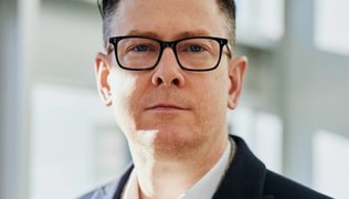
Dr. Michael Chen
Cardiac Surgery
24×7 Video Consultations
25+
Hospitals
2,000+
Providers
60+
Specialities
3M+
Patients
Centres of Excellence
Specialised, multi-disciplinary centres delivering world-class outcomes.
Trauma & Critical Care Centre
The Trauma & Critical Care Centre at SilverOak Health is a major trauma centre providing 24/7 specialist care for patien
Explore →Rheumatology & Autoimmune Diseases Centre
The Rheumatology & Autoimmune Diseases Centre at SilverOak Health provides specialist assessment and management of infla
Explore →Gastroenterology & Hepatology Centre
The Gastroenterology & Hepatology Centre at SilverOak Health provides comprehensive diagnostic and therapeutic services
Explore →Pulmonology & Respiratory Medicine Centre
The Pulmonology & Respiratory Medicine Centre at SilverOak Health provides expert management of acute and chronic lung c
Explore →Diabetes & Endocrinology Centre
The Diabetes & Endocrinology Centre at SilverOak Health provides expert assessment and management of the full spectrum o
Explore →Paediatric Medicine & Surgery Centre
The Paediatric Medicine & Surgery Centre at SilverOak Health provides comprehensive inpatient and outpatient care for ch
Explore →Featured Providers
50 providers across the region
Dr. James Hartwell
Cardiology
Interventional Cardiology
Cardiology
Silvaris Heart Institute — Newark, NJ
22+ years experience
4.9(312)
Dr. Sarah Thompson
Cardiology
Cardiac Electrophysiology
Cardiology
Silvaris Heart Institute — Newark, NJ
17+ years experience
4.8(278)
Dr. Michael Chen
Cardiac Surgery
Cardiac Surgery
Cardiothoracic Surgery
Silvaris Heart Institute — Newark, NJ
25+ years experience
4.9(341)
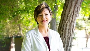
Dr. Emily Rodriguez
Cardiology
Structural Heart Disease
Cardiology
Silvaris Heart & Vascular — Columbus, OH
14+ years experience
4.7(196)
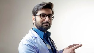
Dr. Robert Williams
Cardiology
Advanced Heart Failure
Advanced Heart Failure
Silvaris Heart Institute — Newark, NJ
20+ years experience
4.8(224)

Dr. Jennifer Davis
Cardiology
Coronary Intervention
Cardiology
Silvaris Heart & Vascular — Columbus, OH
12+ years experience
4.7(168)

Dr. David Kim
Cardiology
Peripheral Vascular Intervention
Cardiology
Silvaris Heart Institute — Newark, NJ
16+ years experience
4.8(207)

Dr. Lisa Anderson
Cardiology
Cardiac Arrhythmia
Cardiology
Silvaris Heart Institute — Newark, NJ
11+ years experience
4.6(143)
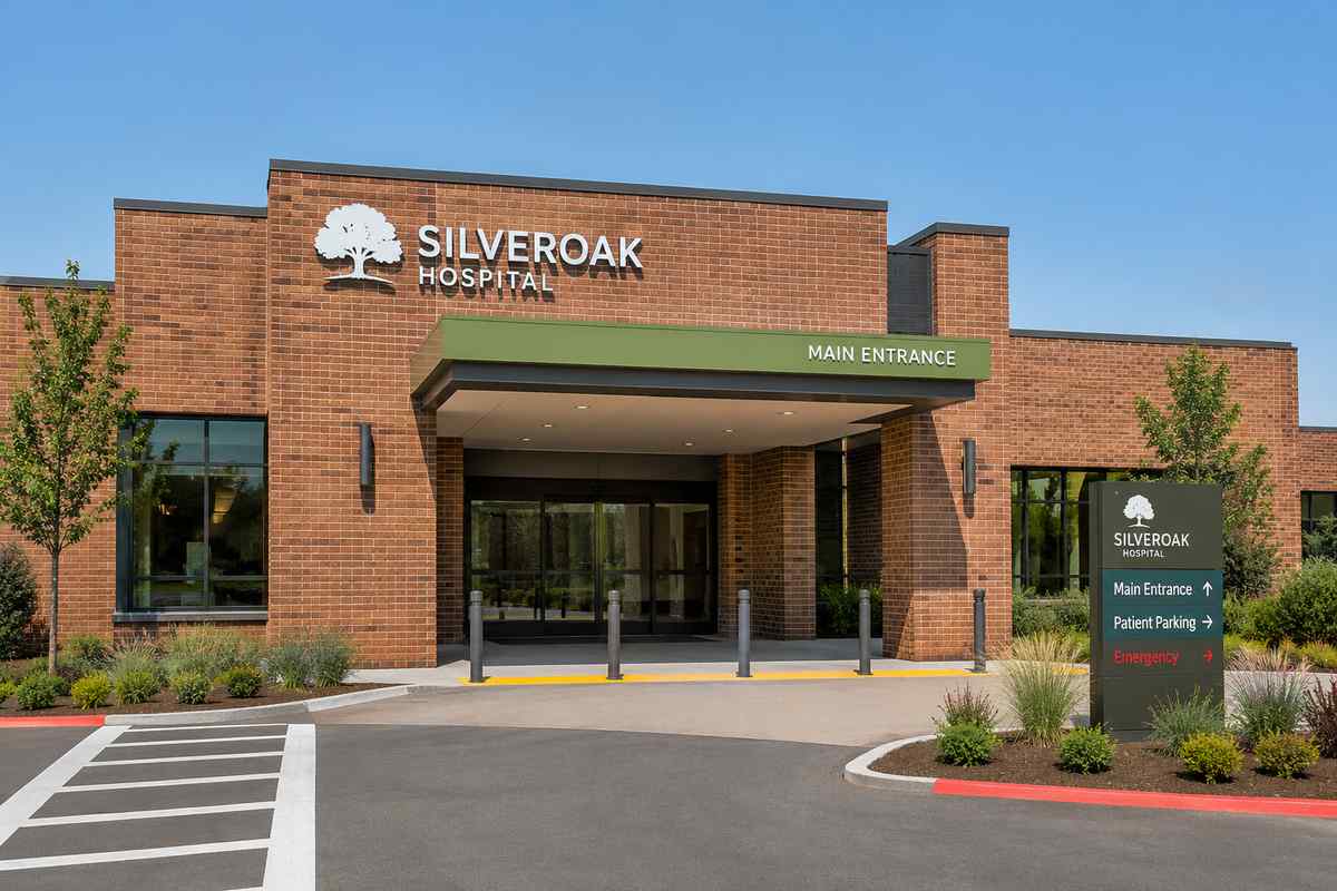
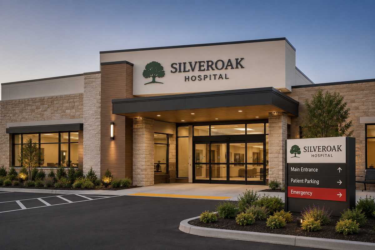
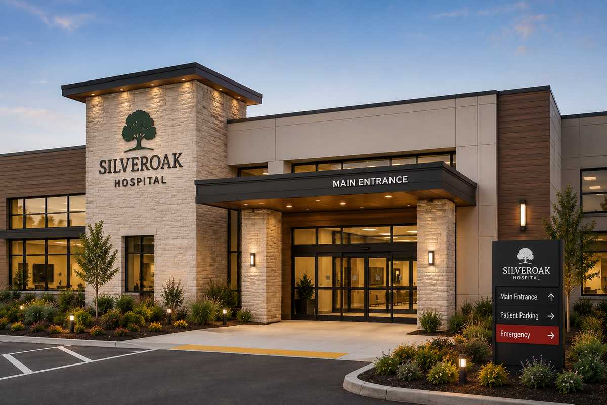
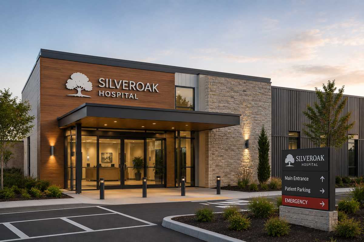
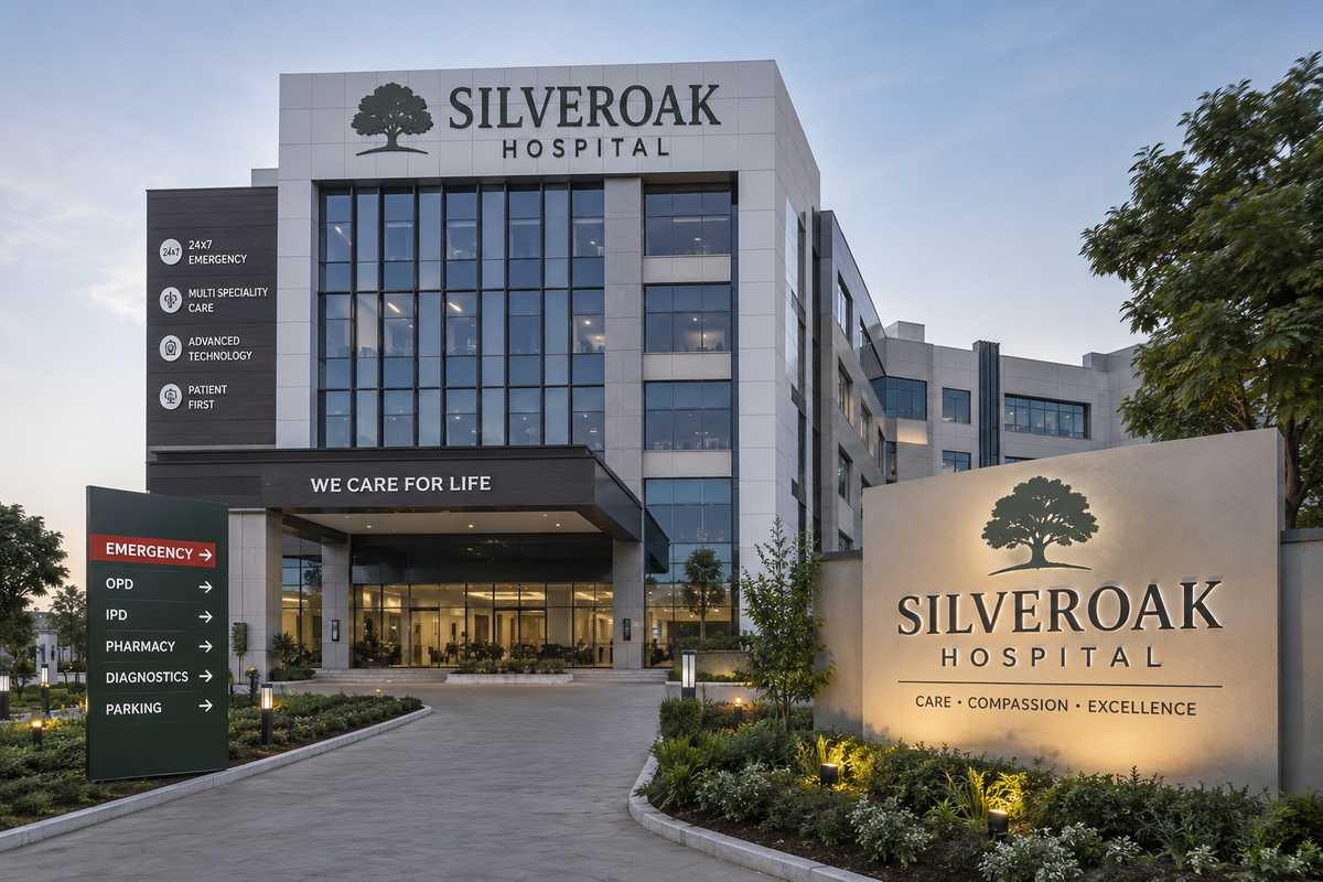
Our Hospitals
Flagship facilities with advanced technology.

Patient Stories
Real patients. Real recoveries.
Rachel Turner was twenty-seven when the road accident that took her colleague's life also shattered her sense of safety in the world. Physically, she recovered — three months of treatment for a fractured pelvis and multiple soft tissue injuries. Psychologically, she did not. In the eighteen months that followed, she stopped driving, stopped sleeping well, and stopped being the person she had been. Flashbacks arrived without warning. Crowds felt dangerous. She quit a job she had loved because the route to work passed the accident site, and she could not make herself drive past it. A friend, recognising the signs, encouraged her to seek help. She was referred to the trauma psychology service at Silvaris Regional Hospital in Dallas. Her initial assessment with the consultant clinical psychologist was the first time Rachel had described the accident and its aftermath to a professional without feeling dismissed or told that time would heal it. 'He took it seriously,' she says. 'He named what was happening — PTSD — and he explained why my brain was responding this way, what the flashbacks were doing neurologically, why the avoidance felt protective but was actually maintaining the problem. Understanding the mechanics of what was happening to me was genuinely helpful. I stopped feeling like I was going mad and started feeling like I had a diagnosable, treatable condition.' A pharmacological review resulted in the prescription of an SSRI to address both the PTSD and comorbid anxiety while she began psychological therapy. Trauma-focused CBT was conducted over sixteen sessions, once weekly. The work was not easy. Trauma-focused therapy requires approaching the memories that the avoidance response has been designed to keep at bay — a process that initially worsened Rachel's distress before improving it. Her psychologist prepared her meticulously for this: explaining the rationale, the expected trajectory, and the fact that feeling worse in the short term was a sign that the work was happening. 'He told me the graph of recovery in PTSD looks like it dips before it rises,' Rachel says. 'And he was right. Around session six or seven I felt the worst I had felt. And then slowly, week by week, the flashbacks became less vivid. The past started to feel less present.' Alongside the psychological therapy, a structured return-to-driving programme was coordinated through a specialist anxiety-management service. Rachel began by sitting in a stationary car, then by short journeys with a therapist in the passenger seat, progressively building her confidence and tolerance for road-related situations. The approach — gradual, graded, always at a pace Rachel could manage — stood in deliberate contrast to the avoidance that had been shrinking her world. Her psychologist and the driving rehabilitation specialist communicated directly, ensuring that the behavioural work was integrated with the therapy. 'Everything was connected,' Rachel says. 'It wasn't a collection of separate interventions — it was a coordinated plan.' Two years after beginning treatment, Rachel drives confidently, has returned to full-time employment in a new role, and has not experienced a flashback in eight months. Her anxiety remains something she manages actively — therapy has given her tools rather than a cure — but it no longer governs her life. She has joined a peer support group for trauma survivors, finding both community and purpose in sharing her experience with others at the beginning of their recovery journeys. 'PTSD stole eighteen months of my life,' she says. 'The team at Silvaris gave me back everything that followed. I am not the person I was before the accident, but I have made peace with that. I am someone who survived, and who knows how to ask for help.'

Rachel Turner
Post-traumatic stress disorder (PTSD) and comorbid generalised anxiety disorder following a road traffic accident in which Rachel sustained serious physical injuries and witnessed the death of a fellow passenger. Rachel presented eighteen months after the accident with intrusive flashbacks, hypervigilance, chronic sleep disturbance, social withdrawal, and avoidance of driving and road-related situations. She underwent a course of trauma-focused cognitive behavioural therapy combined with pharmacotherapy, and a structured return-to-daily-functioning programme.
Joshua Bennett's parents learned that their son had a heart defect before he was born. A foetal echo at twenty-two weeks of pregnancy identified Tetralogy of Fallot — a complex congenital abnormality involving four structural defects in the developing heart. For Joshua's parents, the days between that scan and their first appointment at the paediatric cardiac centre at Silvaris Children's Hospital in Charlotte were among the most frightening of their lives. They arrived at that appointment knowing very little except that their unborn child needed open heart surgery. They left three hours later with something they had not expected: informed hope. The paediatric cardiologist who saw them was exceptional at communicating complex information to terrified, non-medical parents. She used diagrams, models, and plain language to explain exactly what the four components of the defect meant for Joshua's heart function, what would happen at birth, and what the surgical plan would involve. She was honest about the risks — the procedure was major, and no cardiac surgery was without risk — but she contextualised those risks alongside the excellent outcomes that the surgical team had achieved with this specific repair. 'She told us the statistics,' Joshua's mother recalls. 'And then she said: this is the plan, this is the team, and this is what we are going to do together. The word together made everything feel different.' Joshua was born at term, became slightly blue with feeding at four months, and was admitted to hospital at five months for planned surgical repair. The evening before the operation, the surgical team visited the family in their room. The paediatric cardiac surgeon sat on the floor with Joshua in his bouncy chair and spoke directly to the baby — a gesture that moved Joshua's father profoundly. 'He was treating our son as a person, not a case,' he says. The operation lasted six hours. VSD closure and RVOT reconstruction were performed under cardiopulmonary bypass. The surgical team emerged to tell the family that everything had gone according to plan, and that Joshua's heart was beating well. The post-operative days in the paediatric cardiac intensive care unit were difficult. Joshua was on a ventilator overnight, with chest drains and multiple monitoring lines. The nurses in the unit were extraordinary — translating what every number on every monitor meant, preparing the family for what to expect at each stage of recovery, celebrating each milestone openly: the removal of the ventilator, the first time Joshua opened his eyes, the first tentative feed. 'They treated us as partners in Joshua's care from day one,' his mother says. 'They taught us how to read the monitor, what was normal, what to report. They respected our instincts as parents.' Joshua was discharged home twelve days after surgery. Joshua is now four years old. He attends nursery five days a week, runs faster than most of his classmates, and shows no cardiovascular limitations whatsoever. His annual cardiology reviews confirm that the repair is holding well, his right ventricular function is normal, and his scar — a narrow pink line down the centre of his chest — is fading year by year. His parents describe the team at Silvaris Children's Hospital as the reason their son is alive and thriving. 'We were just two terrified people who had been given the worst news of our lives,' his mother says. 'The team made us feel that we were in a place where miracles were considered routine. And for Joshua, it turned out they were.'

Joshua Bennett
Tetralogy of Fallot — a congenital cardiac defect comprising ventricular septal defect, right ventricular outflow tract obstruction, overriding aorta, and right ventricular hypertrophy. Joshua was diagnosed prenatally on foetal echocardiography at twenty-two weeks gestation and was transferred to a tertiary paediatric cardiac centre for planned care. He underwent complete intracardiac repair at five months of age, including VSD closure and right ventricular outflow tract reconstruction.
Dorothy Hayes was seventy-nine and had lived alone since her husband's death three years earlier, managing her household with the stubborn independence that her children both admired and worried about. When her neighbour received no answer at the door and found Dorothy on her kitchen floor — she had fallen reaching for the kettle the previous evening — the scene was frightening. A fractured hip was confirmed in the emergency department. Dorothy was frightened and in pain, and she was also profoundly embarrassed. 'I felt like I had failed somehow,' she says. 'That I had let my age catch up with me.' What she encountered at Silvaris Rehabilitation Center in Portland changed that feeling completely. The orthopaedic and orthogeriatric teams worked in tandem from the moment of her admission. The fracture required surgery within twenty-four hours to reduce the risks of prolonged bed rest, and Dorothy was taken to the operating theatre the following morning. The operation went smoothly. More importantly, a comprehensive geriatric assessment — conducted by the orthogeriatric consultant who reviewed all orthopaedic ward patients — identified several factors that had contributed to Dorothy's fall and would need to be addressed to reduce the risk of future ones: osteoporosis that had never been formally treated, a medication regimen that included a drug known to increase fall risk, and some early memory changes that merited monitoring. 'He looked at the whole picture,' Dorothy says. 'Not just the hip.' Mobilisation began on the day after surgery. The physiotherapy team coaxed Dorothy gently but firmly out of bed and onto her feet — an experience she describes as terrifying and empowering in equal measure. 'I was convinced I would fall again,' she says. 'The physiotherapist held my arm and told me that she would not let that happen, and somehow I believed her.' Progress was incremental but consistent: a frame first, then a stick, then short walks without assistance. An occupational therapist assessed her home before discharge and arranged for a series of adaptations — grab rails, a higher toilet seat, a bath board, removal of the loose rugs that had contributed to her fall. The osteoporosis management was initiated before her discharge: a bisphosphonate prescription, vitamin D and calcium supplementation, and a dexa scan referral to quantify her bone density. The medication that increased fall risk was deprescribed and an alternative found. A falls assessment clinic follow-up was arranged for six weeks post-discharge, where she completed a structured balance and strength programme. Her GP was written to with a detailed summary and clear requests for ongoing monitoring of her bone health and medications. The transition from hospital care to community follow-up was meticulously coordinated, something Dorothy's daughter, who had accompanied her throughout, describes as 'seamless and reassuring.' Six months after her fall, Dorothy is walking with a stick for outdoor distances but independently at home without aids. She attends a weekly strength and balance class at her local community centre, a referral that came via the falls service. Her bone density scan confirmed osteoporosis and her medication is protecting against further bone loss. She has not fallen since. She lives independently, cooks her own meals, and sees her children and grandchildren weekly. 'I thought that fall might be the end of my independent life,' Dorothy says. 'The team at Silvaris made sure it was not. They fixed my hip, but more than that, they fixed the reasons it had broken.'

Dorothy Hayes
Intracapsular fractured neck of femur following a mechanical fall at home. Dorothy, aged seventy-nine, was found on her kitchen floor by a neighbour after lying there for approximately six hours. She was admitted via the emergency department, and plain radiography confirmed a displaced intracapsular fracture of the right femoral neck. She underwent cemented hemiarthroplasty within twenty-two hours of admission. Orthogeriatric review identified osteoporosis, polypharmacy, and cognitive frailty as contributing factors requiring comprehensive post-operative management.
Health Hub
Latest articles, videos and research from our providers.

Understanding Osteoporosis: Prevention, Detection, and Treatment
Osteoporosis — characterised by low bone density and deterioration of bone microarchitecture — is a major public health problem affecting an estimated 10 million Americans, with a further 44 million having low bone density that places them at increased fracture risk. Often called a 'silent disease' because bone loss occurs without pain or obvious symptoms, osteoporosis typically declares itself only when a fracture occurs — frequently at the hip, spine, or wrist. In the United States, osteoporosis causes approximately two million fractures per year, at a direct medical cost exceeding 19 billion dollars annually. Bone is dynamic living tissue constantly being remodelled — old bone is broken down by osteoclast cells and new bone is built by osteoblast cells. Peak bone mass is achieved in the late 20s to early 30s, after which a gradual net decline begins. In women, the accelerated oestrogen loss of menopause — particularly in the first five to ten years — dramatically accelerates bone loss, with women losing up to 20% of bone density in this period. Men lose bone more slowly and develop osteoporosis later, though one in four men over 50 will experience an osteoporotic fracture in their lifetime. Several risk factors beyond sex and age increase osteoporosis risk. A family history of hip fracture, personal history of fragility fracture after age 50, long-term corticosteroid use (one of the most common drug-induced causes), smoking, excessive alcohol (more than three units per day), low body weight or eating disorders, calcium and vitamin D deficiency, prolonged immobility, and secondary causes including rheumatoid arthritis, hyperthyroidism, hyperparathyroidism, coeliac disease, and chronic kidney or liver disease all require heightened vigilance and earlier screening. Diagnosis is made by dual-energy X-ray absorptiometry (DEXA) scan, which measures bone mineral density (BMD) at the lumbar spine and femoral neck and expresses the result as a T-score — the number of standard deviations above or below the mean for a healthy young adult. A T-score of -1.0 to -2.5 indicates osteopenia (low bone mass); -2.5 or below defines osteoporosis; -2.5 or below with one or more fragility fractures defines severe osteoporosis. The FRAX algorithm integrates BMD with clinical risk factors to calculate ten-year fracture probability, guiding treatment decisions more precisely than BMD alone. Prevention is most effective when started early, but it is never too late to intervene. Adequate calcium intake throughout life — 1,000 mg daily for adults under 50, 1,200 mg for women over 50 and men over 70 — provides the raw material for bone mineralisation. Vitamin D 800–1,000 IU daily is essential for calcium absorption and direct bone health. Weight-bearing and resistance exercise — including walking, hiking, dancing, and weightlifting — stimulate osteoblast activity and have the dual benefit of improving balance and reducing fall risk. Smoking cessation and alcohol moderation are modifiable protective measures. Pharmacological treatment is recommended for postmenopausal women and men over 50 with T-score ≤-2.5, or T-score between -1 and -2.5 with a ten-year hip fracture probability ≥3% or major osteoporotic fracture probability ≥20% on FRAX. Oral bisphosphonates — alendronate and risedronate — remain the most widely used first-line agents, reducing vertebral fracture risk by 40–70% and hip fracture risk by 40–50%. For patients unable to tolerate oral bisphosphonates, annual intravenous zoledronic acid is a highly effective alternative requiring only yearly infusion. For patients with severe osteoporosis, multiple fractures, or inadequate response to antiresorptive therapy, anabolic agents that actively build bone offer superior fracture reduction. Teriparatide (recombinant PTH 1-34) and abaloparatide (PTH-related protein analogue) stimulate new bone formation and reduce vertebral fracture risk by up to 65%. Romosozumab, a monoclonal antibody blocking sclerostin, has the dual mechanism of building bone while simultaneously reducing resorption, producing the largest increases in bone density of any available agent. These agents are typically followed by antiresorptive therapy to consolidate gains. Fall prevention is an essential component of osteoporosis management, because fractures require both fragile bone and a fall. Home hazard assessment (removing loose rugs, improving lighting, installing grab rails), balance and strengthening exercises (tai chi has strong evidence in fall prevention), annual vision and hearing checks, medication review to identify drugs contributing to dizziness or postural hypotension, and appropriate footwear all reduce fall risk in the older adult. Multidisciplinary fracture liaison services, which systematically identify and treat patients with fragility fractures, are proven to prevent subsequent fractures — but remain underutilised in many health systems.
Read article →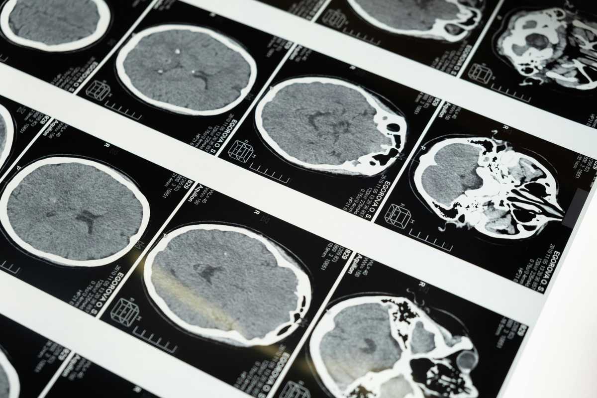
Eye Health: Protecting Your Vision at Every Age
Vision is the sense most people report they would least want to lose — yet eye health is frequently neglected until significant deterioration has already occurred. Over 12 million Americans aged 40 and older have vision impairment, and approximately 1 million are blind. The leading causes of blindness and vision loss in the United States — age-related macular degeneration, diabetic retinopathy, glaucoma, and cataract — are all conditions for which early detection significantly alters outcomes. Regular eye examinations are not a luxury; they are a cornerstone of preventive healthcare. Age-related macular degeneration (AMD) affects the macula — the central portion of the retina responsible for sharp, detailed vision — and is the leading cause of irreversible vision loss in adults over 50. Dry AMD, characterised by the gradual accumulation of drusen deposits and retinal pigment epithelium atrophy, progresses slowly and currently has limited treatment options, though dietary antioxidant supplementation (AREDS2 formula) reduces the risk of progression to advanced disease. Wet AMD — caused by abnormal blood vessel growth beneath the retina — progresses rapidly but can be stabilised with regular intravitreal injections of anti-VEGF agents (ranibizumab, aflibercept, faricimab) if identified early. Diabetic retinopathy is the most common complication of diabetes and the leading cause of new blindness in working-age adults. Chronically elevated blood glucose damages the retinal capillaries, causing leakage, haemorrhage, and eventually the growth of fragile new vessels that can bleed catastrophically or cause traction retinal detachment. Approximately one-third of people with diabetes have some degree of diabetic retinopathy, and many are unaware. Annual retinal photography screening is recommended for all diabetic patients — early laser treatment and anti-VEGF injections prevent severe vision loss in the vast majority when initiated promptly. Glaucoma is characterised by progressive optic nerve damage, usually associated with elevated intraocular pressure, causing irreversible loss of peripheral visual field that advances toward central vision if untreated. It is called the 'silent thief of sight' because most people have no symptoms until significant, permanent visual field loss has already occurred. Early detection through regular intraocular pressure measurement, optic disc assessment, and visual field testing allows treatment with pressure-lowering eye drops, laser therapy, or surgery that effectively halts progression in the majority of patients. Cataracts — clouding of the crystalline lens — are the most common cause of correctable blindness globally, affecting most people to some degree by their 70s. Risk factors include age, UV exposure, diabetes, smoking, prolonged corticosteroid use, and prior eye trauma or surgery. Symptoms include gradually worsening blurred or foggy vision, glare, halos around lights, and difficulty reading or driving at night. Surgical replacement of the cloudy lens with a clear artificial intraocular lens is one of the most commonly performed and safest surgical procedures worldwide, restoring vision in the overwhelming majority of patients. Protecting eye health throughout life begins with UV protection. Wearing sunglasses that block 99–100% of UVA and UVB radiation reduces the risk of cataract, macular degeneration, and photokeratitis. A diet rich in dark leafy vegetables (kale, spinach), coloured fruits, omega-3 fatty acids, and foods containing zinc provides lutein, zeaxanthin, vitamins C and E, and zinc — nutrients that support macular health. Smoking doubles the risk of AMD and significantly accelerates cataract development — cessation is one of the most impactful eye health interventions available. Digital eye strain has become ubiquitous with the proliferation of screens. The 20-20-20 rule — every 20 minutes, look at something 20 feet away for at least 20 seconds — provides regular relief from the sustained near focus that contributes to eye fatigue, headache, and dry eye symptoms. Blinking rate decreases significantly when using screens, compounding dryness; artificial tear drops provide symptomatic relief. Children's excessive screen time has been associated with a dramatic increase in myopia (short-sightedness) globally — outdoor time in natural light appears to be protective. Adults with no risk factors should have a comprehensive eye examination every two years from age 40, and annually from 60. Anyone with diabetes, a family history of glaucoma, previous eye disease, or visual symptoms should be examined annually or more frequently as advised. If you notice sudden floaters or flashes of light, a curtain or shadow across your visual field, or sudden loss of vision — seek emergency ophthalmological assessment immediately. These symptoms may indicate a retinal detachment, which is a surgical emergency with excellent outcomes when treated within 24 hours.
Read article →
The Rise of Robotic Surgery: What Patients Need to Know
Robotic surgery has emerged over the past two decades as one of the most transformative developments in surgical care, bringing a level of precision, visualisation, and control to minimally invasive procedures that was previously unachievable. Since the FDA approval of the da Vinci Surgical System in 2000, robotic-assisted procedures have expanded from urology into gynaecology, general surgery, thoracic surgery, cardiac surgery, colorectal surgery, orthopaedics, and head-and-neck surgery. Understanding what robotic surgery is — and what it is not — helps patients have realistic, informed conversations with their surgical teams. Despite the term, 'robotic surgery' does not involve an autonomous robot operating independently. The surgeon sits at a console several feet from the patient, viewing a high-definition three-dimensional magnified image of the surgical field and controlling robotic arms that hold miniaturised instruments through tiny incisions. The system translates the surgeon's hand movements — filtering out tremor and scaling down the motion — into precise instrument manipulation inside the patient's body. The surgeon is in complete control at every moment; the 'robot' is a sophisticated tool extending human capability. The da Vinci system's EndoWrist instruments can rotate 360 degrees, surpassing the natural range of motion of the human wrist within a constrained body cavity. Combined with the three-dimensional magnified view (up to 10–15x optical magnification), this dexterity enables dissection around delicate structures — the neurovascular bundles in prostatectomy, the recurrent laryngeal nerve in thyroid surgery, or the ureter in hysterectomy — with a precision and safety margin that is genuinely difficult to replicate in conventional laparoscopic surgery. Compared to open surgery, robotic-assisted procedures typically offer several advantages: smaller incisions (typically five to eight millimetre ports), reduced blood loss, lower risk of surgical site infection, shorter hospital stays (often one to two days rather than five to seven for equivalent open procedures), faster return to normal activities and work, and reduced post-operative pain requiring less opioid analgesia. Compared to conventional laparoscopy, the ergonomic advantages reduce surgical fatigue in long complex cases, potentially improving precision consistency throughout a procedure. Robotic surgery is now routinely used for prostatectomy (removal of the prostate for cancer), hysterectomy and myomectomy (uterine fibroid removal), colorectal surgery including hemicolectomy and anterior resection, cholecystectomy, fundoplication for gastro-oesophageal reflux, thyroid and parathyroid surgery, lung resections, and heart valve repair and bypass grafting. In orthopaedics, robotic-arm systems such as Mako assist surgeons in planning and executing knee and hip replacement with sub-millimetre accuracy in implant positioning. Robotic surgery is not suitable for every procedure or every patient. Emergency surgery, procedures requiring large incisions for specimen extraction, and patients with extensive prior abdominal scarring or severe cardiopulmonary disease requiring short operative times may be better served by conventional approaches. The appropriateness of robotic surgery depends on the specific procedure, the patient's anatomy and comorbidities, and critically, the surgeon's experience and training on the robotic platform — outcomes are closely tied to the volume and expertise of the surgical team. Patients considering robotic surgery should ask their surgeon several important questions: How many robotic procedures of this type have you performed? What is your conversion rate to open surgery? Are the outcome data for your robotic cases comparable to published benchmarks? What are the specific advantages and disadvantages of robotic versus conventional approaches for my particular condition? These questions will help you make a genuinely informed decision rather than choosing based solely on the appeal of technology. At SilverOak Health, our robotic surgery programme is led by surgeons with extensive platform-specific training and high procedural volumes, ensuring that patients receive the full benefit of robotic technology. We are committed to offering robotic approaches where evidence supports their use, while being transparent about cases where conventional or laparoscopic surgery remains the better option. Your safety and outcome are always the primary consideration.
Read article →Book your appointment today.
In-person or video — the right provider is a few clicks away.